Real-time analysis of RNA G-quadruplexes in active cells is now possible thanks to a novel microscopic technique, which has implications for the treatment of amyotrophic lateral sclerosis.
Amyotrophic lateral sclerosis (ALS), also referred to as Stephen Hawking’s disease and Lou Gehrig’s disease, is a neurodegenerative condition that causes the body’s muscles to gradually lose control. Over 90% of cases have an unknown cause, but both genetic and environmental factors are thought to be involved. It is currently incurable.
The research teams of Prof. Jerker Widengren at the KTH Royal Institute of Technology in Sweden and Dr. Akira Kitamura at the Faculty of Advanced Life Science at Hokkaido University have created a novel method that can recognize an RNA’s distinctive structure in real time in living cells. In the journal Nucleic Acids Research, the fluorescence-microscopic spectroscopy-based method was described.
“One of the genetic factors that is believed to be involved in the development of ALS is a specific sequence of RNA that forms a four-stranded structure, called a G-quadruplex,” explains Kitamura, first author of the study.
“Normally, these structures regulate the expression of genes. However, a mutation in chromosome 9 in humans results in the formation of G-quadruplexes that may play a role in neurodegenerative diseases including ALS.”
The inability to observe the formation and location of G-quadruplexes in living cells in real time has been one of the biggest obstacles to understanding the precise function of G-quadruplexes in disease. The Kitamura and Widengren groups were successful in creating a straightforward, reliable, and broadly applicable technique that addresses current problems.
The technique tracks a cyanine dye called Alexa Fluor 647 (AF647). When labeled to RNA, the fluorescence blinking state of the dye is altered with formation of the RNA G-quadruplexes. The groups analyzed the AF647-labeled RNA using a microscopy technique called TRAST (TRAnsient STate) monitoring to detect this fluorescence blinking in real time.
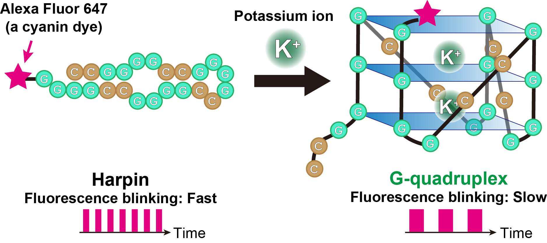
The fluorescent blinking of cyanine dye (Alexa Fluor 647, pink star) bound to RNA changes depending on the structure of the RNA. When the RNA is folded like a hairpin, the fluorescent blinking is fast, and when the RNA switches to a G-quadruplex, the blinking is slow (Akira Kitamura).
“Visually, the time-resolved changes in intensity of fluorescence appear as blinking”, said Kitamura, describing the technique. “In TRAST, we expose cells to a specific pattern of changing light intensities and measure the average intensity of fluorescence emitted from the RNA-bound dye in the cells across specific time intervals. By measuring changes in blinking properties, we can distinguish the structures of RNA within the cell.”
Under controlled laboratory conditions, the team calibrated their experiment and identified precisely which fluorescence blinking represented RNA G-quadruplexes. This information allowed them to use TRAST to locate RNA G-quadruplexes within living cells.
The research presented here demonstrates that cyanine dyes can provide sensitive readout parameters on the RNA G-quadruplex folding states in living cells, even for single cells. The ability to study RNA G-quadruplexes in disease in real time at the intracellular level is consequently made possible. It can also be used to research how proteins in cells fold and misfold.